cardiovascular Crossword
This printable crossword puzzle on the topic of Human Body Systems has 32 clues. Answers range from 3 to 22 letters long. This crossword is also available to download as a Microsoft Word document or a PDF.
Description
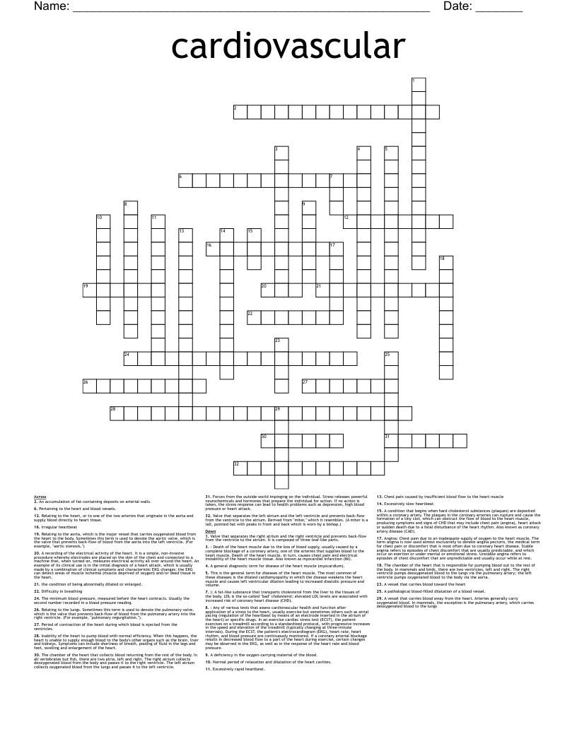
Relating to the aorta, which is the major vessel that carries oxygenated blood from the heart to the body. Sometimes this term is used to denote the aortic valve, which is the valve that prevents back-flow of blood from the aorta into the left ventricle. (For example, "aortic stenosis.")
A vessel that carries blood away from the heart. Arteries generally carry oxygenated blood. In mammals, the exception is the pulmonary artery, which carries deoxygenated blood to the lungs
The chamber of the heart that collects blood returning from the rest of the body. In all vertebrates but fish, there are two atria, left and right. The right atrium collects deoxygenated blood from the body and passes it to the right ventricle. The left atrium collects oxygenated blood from the lungs and passes it to the left ventricle.
Relating to the heart, or to one of the two arteries that originate in the aorta and supply blood directly to heart tissue.
Valve that separates the left atrium and the left ventricle and prevents back-flow from the ventricle to the atrium. Derived from "miter," which it resembles. (A miter is a tall, pointed hat with peaks in front and back which is worn by a bishop.)
Relating to the lungs. Sometimes this term is used to denote the pulmonary valve, which is the valve that prevents back-flow of blood from the pulmonary artery into the right ventricle. (For example, "pulmonary regurgitation.").
Valve that separates the right atrium and the right ventricle and prevents back-flow from the ventricle to the atrium. It is composed of three leaf-like parts.
A vessel that carries blood toward the heart
The chamber of the heart that is responsible for pumping blood out to the rest of the body. In mammals and birds, there are two ventricles, left and right. The right ventricle pumps deoxygenated blood to the lungs via the pulmonary artery; the left ventricle pumps oxygenated blood to the body via the aorta.
A deficiency in the oxygen-carrying material of the blood.
A pathological blood-filled dilatation of a blood vessel.
Chest pain caused by insufficient blood flow to the heart muscle
Irregular heartbeat
An accumulation of fat-containing deposits on arterial walls.
Excessively slow heartbeat
Normal period of relaxation and dilatation of the heart cavities.
the condition of being abnormally dilated or enlarged.
Difficulty in breathing
This is the general term for diseases of the heart muscle. The most common of these diseases is the dilated cardiomyopathy in which the disease weakens the heart muscle and causes left ventricular dilation leading to increased diastolic pressure and volume.
Period of contraction of the heart during which blood is ejected from the ventricles.
Excessively rapid heartbeat.
Angina: Chest pain due to an inadequate supply of oxygen to the heart muscle. The term angina is now used almost exclusively to denote angina pectoris, the medical term for chest pain or discomfort that is most often due to coronary heart disease. Stable angina refers to episodes of chest discomfort that are usually predictable, and which occur on exertion or under mental or emotional stress. Unstable angina refers to episodes of chest discomfort that are unpredictable and usually occur while at rest.
A general diagnostic term for disease of the heart muscle (myocardium).
Pertaining to the heart and blood vessels.
Inability of the heart to pump blood with normal efficiency. When this happens, the heart is unable to supply enough blood to the body's other organs such as the brain, liver and kidneys. Symptoms can include shortness of breath, pooling of fluid in the legs and feet, swelling and enlargement of the heart.
A condition that begins when hard cholesterol substances (plaques) are deposited within a coronary artery. The plaques in the coronary arteries can rupture and cause the formation of a tiny clot, which can obstruct the flow of blood to the heart muscle, producing symptoms and signs of CHD that may include chest pain (angina), heart attack or sudden death due to a fatal disturbance of the heart rhythm. Also known as coronary artery disease (CAD).
The minimum blood pressure, measured before the heart contracts. Usually the second number recorded in a blood pressure reading.
A recording of the electrical activity of the heart. It is a simple, non-invasive procedure whereby electrodes are placed on the skin of the chest and connected to a machine that, when turned on, measures electrical activity all over around the heart. An example of its clinical use is in the initial diagnosis of a heart attack, which is usually made by a combination of clinical symptoms and characteristic EKG changes; the EKG can detect areas of muscle ischemia (muscle deprived of oxygen) and/or dead tissue in the heart.
: Death of the heart muscle due to the loss of blood supply, usually caused by a complete blockage of a coronary artery, one of the arteries that supplies blood to the heart muscle. Death of the heart muscle, in turn, causes chest pain and electrical instability of the heart muscle tissue. Also known as myocardial infarction (MI).
): A fat-like substance that transports cholesterol from the liver to the tissues of the body. LDL is the so-called "bad" cholesterol; elevated LDL levels are associated with increased risk of coronary heart disease (CHD).
Forces from the outside world impinging on the individual. Stress releases powerful neurochemicals and hormones that prepare the individual for action. If no action is taken, the stress response can lead to health problems such as depression, high blood pressure or heart attack.
: Any of various tests that assess cardiovascular health and function after application of a stress to the heart, usually exercise but sometimes others such as atrial pacing (regulation of the heartbeat by means of an electrode inserted in the atrium of the heart) or specific drugs. In an exercise cardiac stress test (ECST), the patient exercises on a treadmill according to a standardized protocol, with progressive increases in the speed and elevation of the treadmill (typically changing at three-minute intervals). During the ECST, the patient's electrocardiogram (EKG), heart rate, heart rhythm, and blood pressure are continuously monitored. If a coronary arterial blockage results in decreased blood flow to a part of the heart during exercise, certain changes may be observed in the EKG, as well as in the response of the heart rate and blood pressure.