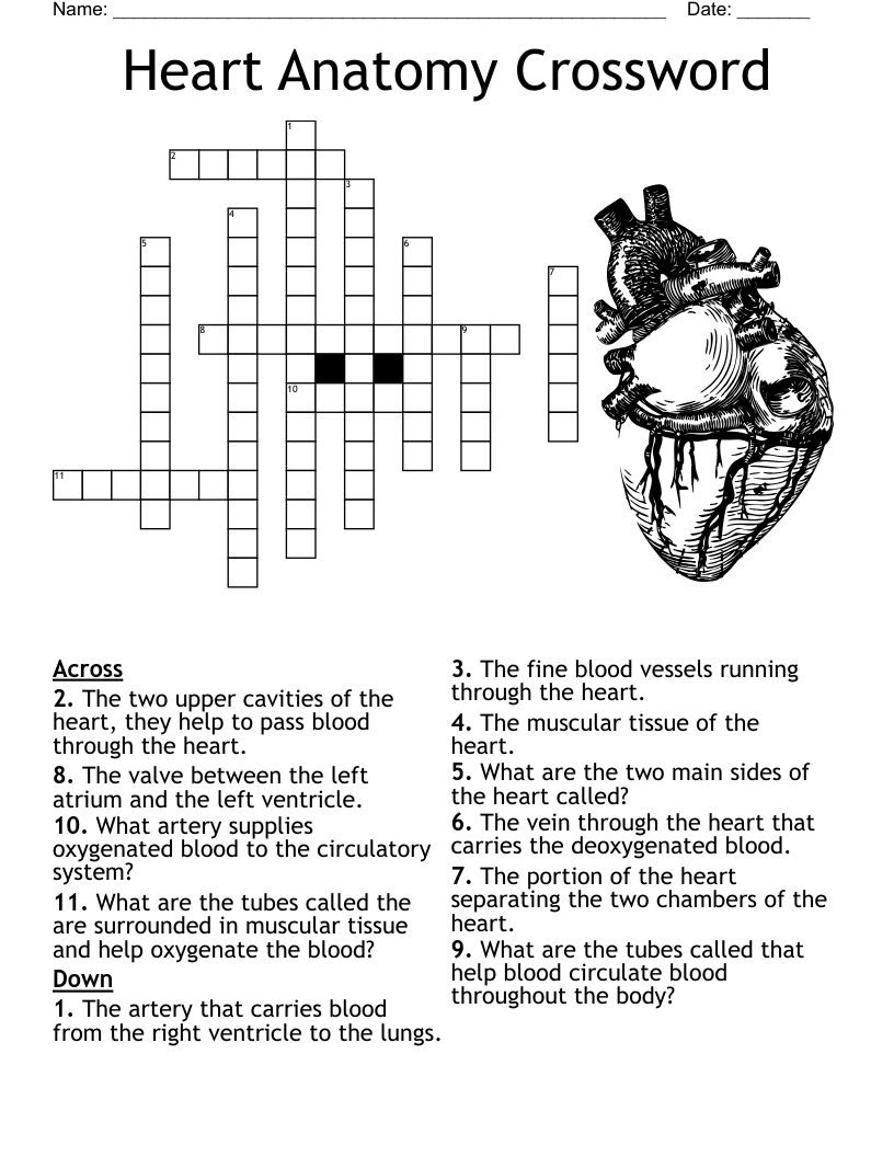
It is also known as the “transportation” system of the body
Smooth layer, Lines the interior, and valves are made from this layer
Muscle layer and Thickest layer
Thin, outermost layer. Joins with lining outside the heart to form the Pericardium
Separates the left and right heart
top part of the septum
bottom part of the septum
receives blood from the superior and inferior vena cava
receives blood from the right atrium, pumps blood to the lungs
receives oxygenated blood from the pulmonary veins
receives blood from the left atrium, pumps oxygenated blood to the rest of the body, strongest chamber
Are important to control the flow of blood from one chamber of the heart to another and allows blood to flow in only one direction
threads, keep valve flaps from flipping up into the atria
opening between right atria and right ventricle
opening between right ventricle and pulmonary artery
opening between left atrium and left ventricle
located between left ventricle and aorta
needs blood supply, blood supplied through the right and left coronary arteries
collects venous blood from the heart and empties into the right atrium
Another name for tricuspid valve
Another name for the pulmonary valve
Another name for the Bicuspid/Mitral valve
Main artery of the body
Carries blood from upper body
Carries blood from lower body
Left lower chamber of heart that receives blood from the left atrium
Deoxygenated blood enters from Inferior Vena Cava to this chamber
Carries high O2 blood towards the heart
The Semilunar Valve seperating the Aorta from the Left Ventricle that prevents bloodflow backwards
Acts as a holding chamber for blood returning from lungs and acts as pump to transport blood
Right side valve between Right Atrium and the Right Ventricle
Pumps Low O2 blood out of the heart
The valve between the Left Atrium ad Left Ventricle
Between Left Ventricle and Aorta
Right side lower chamber that receives blood from Right Atrium
Function is to make sure blood is flowing how it should
Found on both sides of the heart
Double walled sac that encloses the heart
Outer layer of the heart that is continuous with the pericardium
Forms the bulk of the heart and is composed of mostly cardiac muscle
Glistening white sheet of endothelium that lines the inside of heart chambers, 3rd layer of heart
Chamber of heart that deoxygenated blood from systemic circulation enters
AV valve that separates right atrium and right ventricle
Lower chamber of heart that forces blood into pulmonary circulation
Blood flows through the ________________ valve into the pulmonary trunk.
Blood returning from the lungs flows into this chamber
Two-cusped valve between left atrium and ventricle
Left chamber that pumps blood into systemic circulation
The left ventricle pumps blood through this valve into the aortic trunk
This vessel branches to send blood to both lungs
Large branching vessel that is the beginning of systemic circulation
The thicker myocardium of the ventricles that exherts a greater force
The thinner myocardium of the atria has bundles of muscles called _____________.
Dense network of connective tissue fiber that reinforces myocardium and anchors cardiac muscle fibers
Vessels that carry blood away from heart
Vessels that carry blood back to the heart
small vessels in which blood and tissue cells exchange oxygen and carbon dioxide
Functional blood supply of the heart
the main artery of the body, supplying oxygenated blood to the circulatory system. In humans it passes over the heart from the left ventricle and runs down in front of the backbone.
is a valve in the human heart between the left ventricle and the aorta. It is one of the two semilunar valves of the heart, the other being the pulmonary valve.
the mitral valve(bicuspid valve), and the tricuspid valve, which are between the upper chambers (atria) and the lower chambers (ventricles).
is simply the amount of blood pumped by the heart per minute. Necessarily, the cardiac output is the product of the heart rate, which is the number of beats per minute, and the stroke volume, which is amount pumped per beat.
the phase of the heartbeat when the heart muscle relaxes and allows the chambers to fill with blood.
the thin, smooth membrane that lines the inside of the chambers of the heart and forms the surface of the valves.
a serous membrane that forms the innermost layer of the pericardium and the outer surface of the heart.
a large vein carrying deoxygenated blood into the heart. There are two in humans, the inferior vena cava (carrying blood from the lower body) and the superior vena cava (carrying blood from the head, arms, and upper body).
is the wall of tissue that separates the right and left atria of the heart.
is the stout wall separating the lower chambers (the ventricles) of the heart from one another.
is a valve with two flaps in the heart, that lies between the left atrium and the left ventricle.
the muscular tissue of the heart.
are muscles located in the ventricles of the heart.
each of a pair of valves in the heart, at the bases of the aorta and the pulmonary artery, consisting of three cusps or flaps that prevent the flow of blood back into the heart.
is the volume of blood pumped from the left ventricle per beat.
Carries blood away from the heart
Carries blood towards the heart
The largest artery in the body
Join together to form venules
_____ in veins ensure that blood travels only in the direction of the heart
Largest vein in the body
Veins collect _______blood from the tissues
The vena cava empty into the ____ ____
Blood passes through the tricuspid valve into the right ______
During ______, right ventricle constricts and tricuspid valve closes
Blood is ejected from the right ventricle through the _____ valve into the pulmonary arteries
Pulmonary veins bring blood into the ___ ___
Blood flows from left atrium through the ______ valve and into the left ventricle
Type of muscle in the wall of most blood vessels
The aortic and pulmonic valves are called _____ valves
Vein that lies along the medial aspect of the hind limb between the groin and tarsal joint
The vein that is located on the front of the foreleg
superior vena cave
inferior vena cave
pulmonary Artery
pulmonary valve
tricuspid valve
right ventricle
pulmonary vein
left ventricle
aortic valve
mitral valve
right atrium
pericardium
left atrium
Aorta
Heart
septum
tricuspid valve
pulmonary valve
mitral valve
aortic valve
aorta
myocardium
endocardium
mediastinum
pericardium
arteries
veins
capillaries
thoracic cavity
vasoconstriction
vasodilation
vena cava
SA node
systole
diastole
atrium
ventricles
apex
leaflets
means pertaining to the heart and blood vessels.
is a hollow, muscular organ located in the thoracic cavity, between the lungs.
is the double-walled membranous sac that encloses the heart.
is the external layer of the heart and the inner layer of the pericardium.
is the middle and thickest of the heart's three layers.
which consists of epithelial tissue, is the inner lining of the heart.
which supply oxygen-rich blood to the myocardium.
are the two upper chambers of the heart, and these chambers are divided by the interatrial septum.
are two lower chambers of the heart, and these chambers are divided by the inter ventricular septum.
controls the opening between the right atrium and the right ventricle.
is located between the right ventricle and the pulmonary artery.
is located between the left atrium and left ventricle.
is located between the left ventricle and the aorta.
is the flow of blood only between the hearts and lungs.
carry deoxygenated blood out of the right ventricle and into the lungs.
carry the oxygenated blood from the lungs into the left atrium of the heart.
includes the flow of blood to all parts of the body except the lungs.
is the ability to pump blood effectively throughout the body; the contraction and relaxation (beating) of the heart must occur in exactly the correct sequence.
is located in the posterior wall of the right atrium near the entrance of the superior vena cava.
is located on the floor of the right atrium near the interatrial septum.
is a group of fibers located within the inter ventricular septum.
are specialized conductive fibers located within the walls of the ventricles.
are large blood vessels that carry blood away from the heart to all regions of the body.
is the largest blood vessels in the body.
are the major arteries that carry blood upward to the head.
are the smaller, thinner branches of the arteries that deliver blood to the capillaries.
which are only one epithelial cell in thickness, are the smallest blood vessels in the body.
form a low-pressure collecting system to return oxygen-poor blood to the heart.
are the smallest veins that join to form the larger veins.
are the two largest veins in the body.
Returns deoxyginated blood back to the heart
The artery that carries blood from the heart to the body
Valve between right atrium and right ventricle
Valve opening and closing to the aorta
Top two chambers of the heart
Vein returning oxygenated blood from the lungs to the heart
Carries deoxygenated blood from the right ventricle through the pulmonary _________ .
Also known as the mitral valve
Two lower chambers of the heart that expand during diastole
Valve opening for deoxygentated blood to flow to the lungs from the right ventricle
Heart is made of
Hearts natural pacemaker
Another term for Bicuspid valve
These carry blood back to the heart
Carries deoxygenated blood to the right atrium
Carries deoxygenated blood from lower part of the body to the Right Atrium
Receives deoxygenated blood.
Oxygenated blood enters this chamber first.
This seperates the right and left side of the heart
Largest artery in the body
"heart strings"
Prevents back flow of blood into the right atrium
Pumping chambers of the heart.
Blood vessel carries blood from the heart to the lungs to receive oxygen.
These veins carry oxygenated blood and drain into the left atrium.
The heart has _ _ _ _ chambers
The movement of blood through the heart and body is called
Heart sounds are produced by the closing of the ___
This organ removes waste from blood
Heart muscle relaxes
The Heart is made of